Introductory Biomechanics From Cells to Organisms
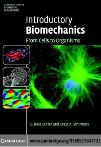
The cover contains images that together represent the broad scope of modern
biomechanics. The figures are as follows:
Main image: A fluorescent immunohistochemical image of an endothelial cell
isolated from the surface of a pig aortic heart valve and grown in culture.Within
the cell, the nucleus is stained blue and vimentin filaments are stained green.
Vimentin is an intermediate filament protein of the cellular cytoskeleton that
plays an important role in cellular mechanics.
Left top: An intermediate stage from a simulation of the forced unfolding of
repeats 4 and 5 of chainAof the protein filamin. Filamin is an actin cross-linking
protein and therefore plays a role in the biomechanics of the cytoskeleton. The
simulation was based on the crystal structure of part of filamin [1], and was
carried out in NAMD [2] and visualized using the VMD package [3]. (Image
courtesy of Mr. Blake Charlebois.)
Left middle: A sketch by the Swiss anatomist Hermann von Meyer of the
orientation of trabecular bone in the proximal human femur. This sketch was
accompanied in the original article by a sketch of the principal stress trajectories
in a crane having a shape similar to the femur. Together these sketches are
believed to have inspired “Wolff’s Law” of bone remodelling. From [4].
Left lower: The distribution of mass transfer rates from flowing blood to cultured
vascular endothelial cells. The contoured quantity (the Sherwood number) was
computed by first measuring the topography of the endothelial cells using atomic
force microscopy and then solving the convection-diffusion equation in the
blood flowing over the cells. Mass transfer from blood to endothelial cells is
important in cell-cell signalling. (Image courtesy of Mr. Ji Zhang.)